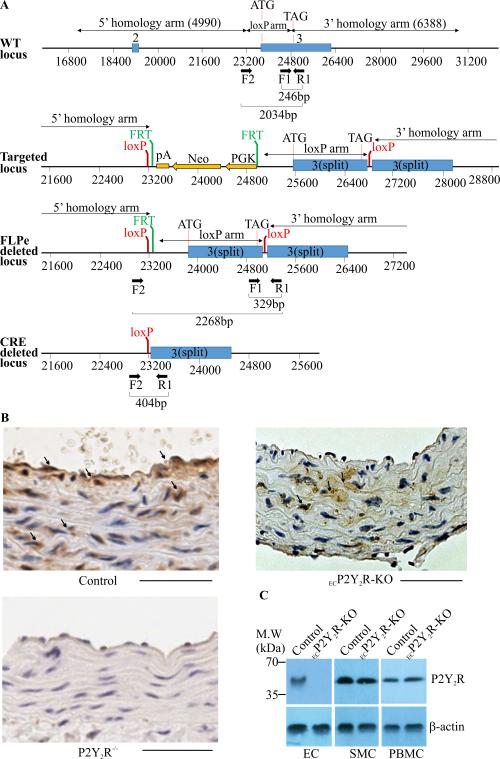
Figure 1.
Generation of ECP2Y2R-KO mice. A. The wild type mouse P2Y2R locus is indicated in the upper panel. The positions of the 5’, 3’ and loxP homology arms used to generating the targeting vector are indicated. Exons 2 and 3 of the gene are indicate by blue boxes. The positions of primers used for genotyping are also indicated. Schematics of the targeted locus, the FLPe deleted locus, in which the neo cassette was removed and the final cre deleted or knockout locus are also shown. Cre-mediated deletion results in excision of the entire coding region of the P2Y2R gene. B, Immunostaining of P2Y2R in cross sections of aortic vessels in control, ECP2Y2R-KO and P2Y2R−/− (total body P2Y2R knock out) mice. P2Y2R is expressed in both EC and SMC in aorta of control mice whereas in ECP2Y2R-KO mice expression was only detected in SMC. P2Y2R staining was absent from EC and SMC in the aorta of P2Y2R−/−mice. Scale bar represents 50μm. C, Western blot analysis of P2Y2R expression in primary cultured EC, SMC and PBMC from control and ECP2Y2R-KO mice. Data shown are representative of experiments performed from 4 mice for each genotype.

