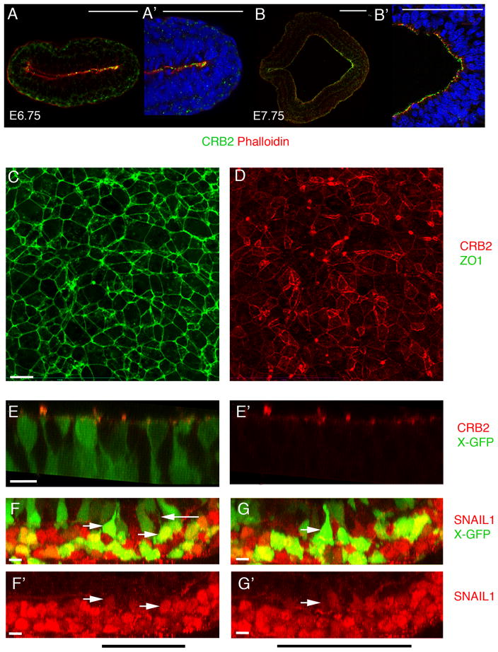
Figure 5. CRB2 enrichment at the primitive streak.
(A, B) Double staining of CRB2 and phalloidin at early primitive streak stages, posterior to the right. (A, A′) At E6.75, CRB2 is enriched in the posterior epiblast, including the region of the primitive streak. Posterior epiblast from A is magnified in A′. (B, B′). At E7.75 CRB2 is expressed in both the anterior presumptive neural epithelium and the epiblast and is enriched in the posterior epiblast. (B′) Higher magnification of streak region. (C, C′) En face view of the apical surface of E8.5 posterior epiblast cells; proximal is up. Extended projection view of the same region of the primitive streak stained for ZO1(C) and CRB2 (D). ZO1 is expressed at uniform levels on all cell edges, whereas the level of expression of CRB2 is highly variable between cells and at different edges of the same cell. Neither Crumbs2 RNA nor CRB2 protein was detected in the wild-type endoderm or mesoderm (Fig. 5A, A′; Supplementary Fig. 1). (E, E′) 3D reconstruction from whole mount immunostaining for CRB2 in wild-type E8.0 embryos that express the X-linked GFP transgene, highlight the shape of individual cells. Apically constricted cells have high levels of apical CRB2. (F–G′) 3D reconstruction from whole mount immunostaining for SNAIL1 in wild type X-GFP embryos at E8.0. SNAIL1 is detectable in the basal nuclei of apically constricted cells in the epiblast layer of the wild-type streak. Black bar indicates the position of the streak. Cells of the wild-type mesoderm layer (bottom) are strongly Snail1+ (red), whereas some apically constricted GFP+ cells of the epiblast layer (top) have detectable levels of Snail1 in their basally-positioned nuclei (rightward arrows). Note the thin GFP+ cytoplasmic connection to the apical surface of the Snail+ cell in (F) (leftward arrow). Scale bars A, B, 100 μm; C–G′, 10 μm. Each immunostaining experiment contained at least 5–6 wild type embryos with and without X-GFP each. The images shown here are representative. This staining was repeated a minimum of three independent times to ensure reproducibility.

