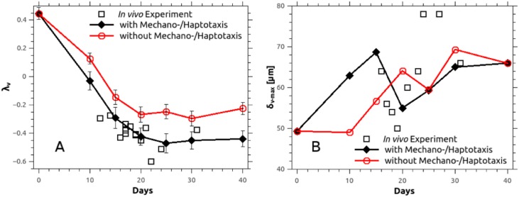
Fig 8. History plots of the parameters characterising the morphology of the microvascular tree.
Numerically predicted parameters λv and δv-max with respect to time, compared to in-vivo measurements in murine mammary carcinoma (MCaIV-type) [22]. A: The geometrical exponent, λv, is obtained after linear regression on the pair of data: frequency of voxels versus the distance to nearest vessel (δv) [65] while the vertical bars denote standard deviation of the mean.

