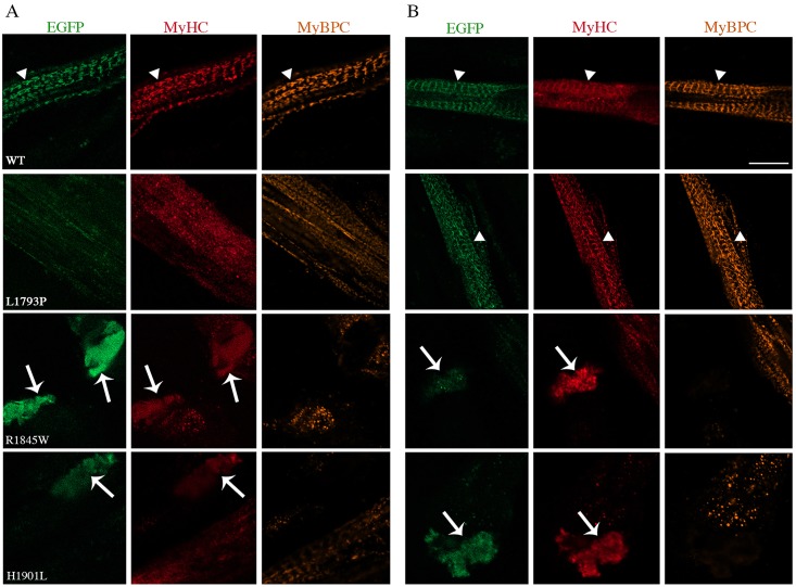
Fig 2. Co-immunofluorescence analysis of 1- and 3-day differentiated myotubes.
Double immunostaining was performed for MyHC (red) and MyBPC (orange) for 1-day (A) and 3-day (B) differentiated myotubes. The repetitive well-structured sarcomere can be seen clearly in 1- and 3-day differentiated myotubes transfected with WT and in 3-day differentiated myotubes with L1793P mutant EGFP-tagged slow/ß-cardiac MyHC constructs (arrowheads). Cells transfected with WT and L1793P mutant were co-immunostained for MyHC and MyBPC. Arrows indicate the accumulation of myosin in 1- and 3-day differentiated myotubes transfected with E1845W and H1901L mutants. Note the myosin accumulations are devoid of MyBPC staining. The bars represent 10 μm.

