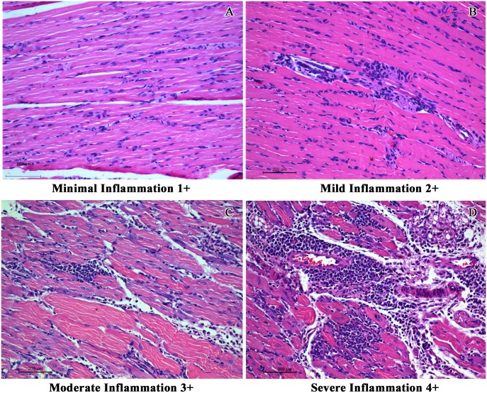
Fig 6. Representative images used for determining the overall inflammation score used in the study.
Inflammation was scored in a scale of 1+ to 4+. A) Minimal inflammation (1+) characterized by sparse inflammatory infiltrate spreading along the interstitium and prominent muscle cell nuclei as a reparative response. B) Mild inflammation (2+) with lymphohistiocytic infiltration. C) Moderate inflammation (3+) with diffuse and focal aggregates of lymphohistiocytic cells spreading between the muscle fibres. Bluish muscle fibres in the section represents the regenerative muscle fibres in response to myositis. D) Severe inflammation (4+) with admixture of lymphohistiocytic and neutrophilic cell infiltrate with myonecrosis and regenerative activity of the muscle fibres. (Scale bar -200μM, H & E stain).

