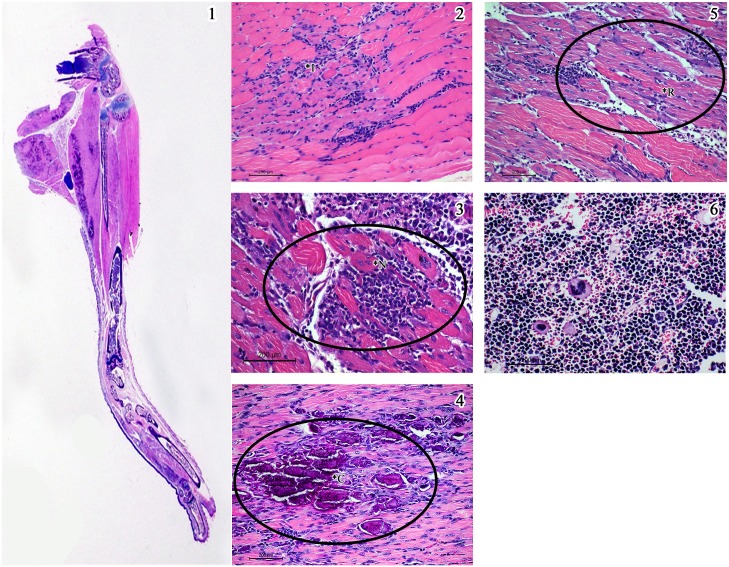
Fig 7. Salient histopathological features in the lower limb in mice injected with CHIKV (Group 1).
1) Whole mount section of the lower limb showing bluish patches of reactive calcification along the margins of the thigh muscle (x 2. H & E stain). 2) Reveals features of myositis with inflammatory infiltrate in Panel (*I). 3) Muscle necrosis with dense inflammatory infiltrate spreading in the endomysium (*N). 4) Mineralization of the necrosed tissue visible as dark blue flakes (*C). 5) Regeneration of the muscle fibres with the bluish pink colour (*R). 6) Hyperplasia of the bone marrow with prominent megakaryocytes. (Scale bar -200μM, H & E stain).

