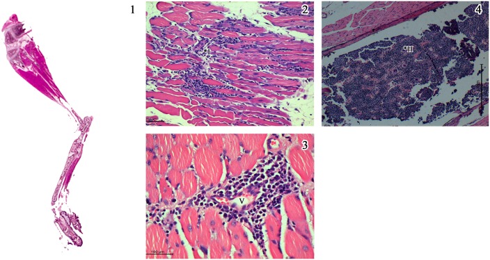
Fig 8. Salient histopathological features in lower limbs in mice injected with CHIKV specific peptides A & B (Group 4 & 5).
1) Whole mount section of lower limb of mice (x 2. H & E stain) with inflammation in the thigh muscle. 2) Endomysium is inflitrated by moderate degree of lymphohistiocytic cells (*I) and the muscle cells undergoing necrosis and atrophy. 3) Higher magnification revealing perivascular infiltration (*V) by lymphoplasmocytic cells and occasional polymorphs representing venulitis (Scale bar -100μM). 4) Marrow space of the femur filled with haemopoietic cells, suggesting marrow hyperplasia (*H). ((Scale bar -200μM, H & E stain).

