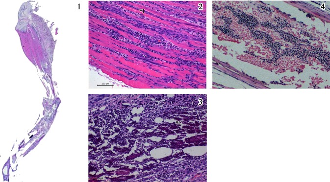
Fig 9. Salient histopathological features noted in H & E stained sections of lower limbs in mice injected with CHIKV followed by specific peptides A & B (Group 7 & 8).
1) Whole mount section of lower limb of the mice showing inflammatory infiltrate in the thigh muscle and extending along the tendon down (x 2. H & E stain). 2) Variable (dense at places) inflammation (*I) between the muscles fibres widening endomyseal space. Occasional bluish regenerating fibres are seen. 3) Dense reactive mineralisation of the necrotic fibres (*C) seen as bluish aggregates admixed with inflammation. 4) Marrow space of the thigh bone showing mild marrow haemopoietic cell hyperplasia. (Scale bar -200μM, H & E stain).

