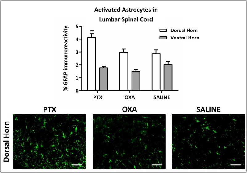
Fig 7. Chemotherapy-induced astrocyte activation in the spinal cord dorsal horn.
GFAP immunohistochemistry in L3-L5 spinal cord was carried out on day 13 post 1st injection of paclitaxel (PTX), oxaliplatin (OXA) or saline (control). Column graph showing the percentage of GFAP immunoreactivity in spinal cord dorsal and ventral horns, and representative images depicting GFAP immunoreactivity in the dorsal horn. GFAP immunoreactivity was significantly higher in the dorsal horn of PTX-treated mice compared with saline controls (**P<0.01), with no significant changes in the ventral horn. Scale bar = 50μm. n = 4, one-way ANOVA followed by Bonferroni's multiple comparison’s test. Data are expressed as mean±SEM.

