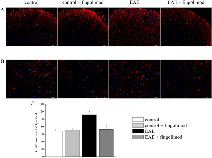
Fig 8. Effects of in vivo prophylactic fingolimod on astrocytes in the spinal cord of EAE mice at the acute stage of disease.
On day 21 post EAE induction, tissue sections were immuno-stained with the anti-GFAP antibody (red) to recognize astrocytes and with DAPI (blue) to identify cell nuclei. (A) 10X: Low-magnification image of spinal cord sections. (B) 20X: High-magnification image of the spinal cord sections. (C) Quantitative evaluation of the number of GFAP-positive cells/Optic field in the spinal cord of mice of each treatment-group. * p < 0.05 versus all other groups; ° p < 0.05 versus untreated EAE mice.

