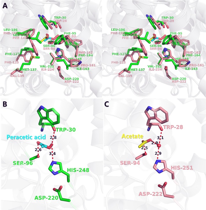
Fig 4. Structural comparisons of ligand-binding site between peracetate-bound EaEST and acetate-bound PfEST.
(A) Stereo view of the superimposed structure of peracetate (cyan)-bound EaEST (green) and acetate (yellow)-bound PfEST (PDB code 3HI4, acetate-bound form, salmon). The residues comprising the active and ligand-binding sites are shown in a stick representation. (B) Peracetate-binding mode and its interactions in EaEST structure. (C) Acetate-binding mode and its interactions in PfEST structure.

