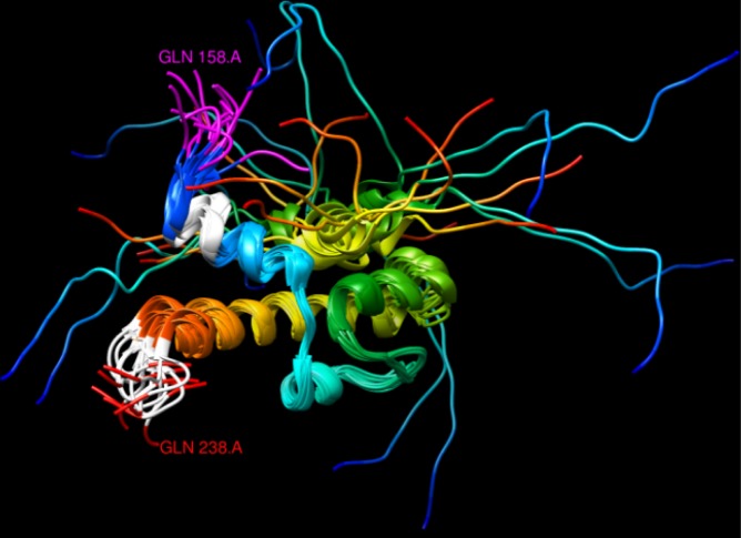
Fig 3. NMR structure of yeast protein Gal11 (PDB:2LPB), residues 158–238.
One 4/4 and another 6/8 polyQ regions are coloured in white (top and bottom, respectively), while the last amino acids of a 12/12 polyQ are coloured in pink. The protein structure is shown with a ribbon representation and non polyQ regions were coloured from blue to red according to the sequence position from N- to C-terminal, respectively. The overlapping structures represent an ensemble of models from the nuclear magnetic resonance (NMR) spectra of the protein in solution.

