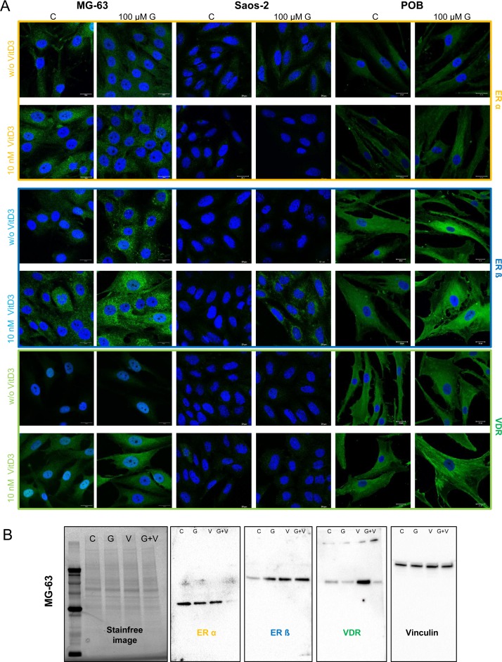
Fig 2. Immunofluorescence staining of ERα, ERß, and VDR in MG-63, Saos-2 and primary osteoblasts (POB) after 48 h exposure to the vehicle control (C), 100 μM genistein (G), 10 nM calcitriol (VitD3) or the simultaneous application of genistein and calcitriol.
Receptor expression was secondarily labeled with Alexa488 (green). All samples were counterstained with DAPI to label the cell nucleus (blue). ERß and VDR expression is highly increased in MG-63 and POB cells after combined treatment with genistein and calcitriol. n = 3.

