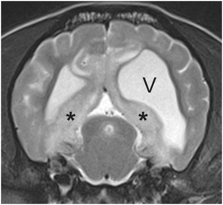
Fig 1. 3T T2W axial MR image at the level of the caudal colliculus illustrating the typical hyperintensity (*) that is often prominent adjacent to the ventricles in cases of MUO.
This dog also exhibits ventricular asymmetry (‘V’ indicates left ventricle), which is a common incidental finding in small breed dogs (this was a pug).

