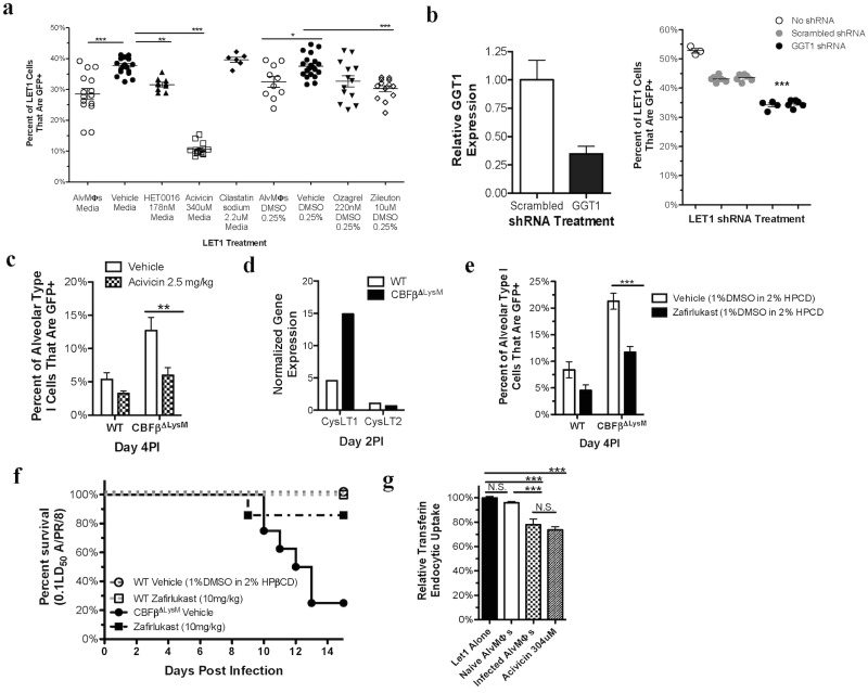
Fig 6. Inhibition of the 5-LOX pathway or blockade CysLT1 renders T1AECs resistant to IAV infection.
a) Infectivity of LET1 cells infected with NS1-GFP A/PR/8 in the presence of the specified treatment. b) Lentivirus shRNA knockdown of GGT1 (left panel) and the impact LET1 cell infectivity (right). c) WT and CBFβΔLysM mice were infected i.n. with NS1-GFP A/PR/8 and treated i.n. with 2.5mg/kg of Acivicin or vehicle at 5 and 29hours post infection. Infection of T1AECs (left) and conducting airway epithelial cells (right) was analyzed at day 4 PI. d) Expression of the CysLT1 and CysLT2 receptors in sorted T1AECs from WT and CBFβΔLysM at day 2PI as determined by RNAseq. e) Day 4 T1AEC infectivity in WT and CBFβΔLysM mice that were infected i.n. with NS1-GFP A/PR/8 and. f) CBFβΔLysM mice that were infected i.n. with 0.1LD50 of A/PR/8 and treated i.p. with 10mg/kg of Zafirlukast or vehicle every 24 hours starting at 5 hours PI until day 3 PI. g) Relative fluorescence of pHrodo Red labeled transferrin that has been taken up via the endocytic route by LET1 cells in the presence of different treatments. For in vitro analyses, data were pooled from, or is representative of, a minimum of 3 experiments with each dot representing 2-pooled wells from a 24well plate. c and e) data was pooled 3 experiments for a total of 4–6 mice for each treatment and genotype. Error bars are standard error mean. Statistical analysis is a either a 2-way ANOVA, 1-way ANOVA, or a two-tailed non-paired students t test. * indicates P< .05, ** for P < .001 and *** for P < .001.

