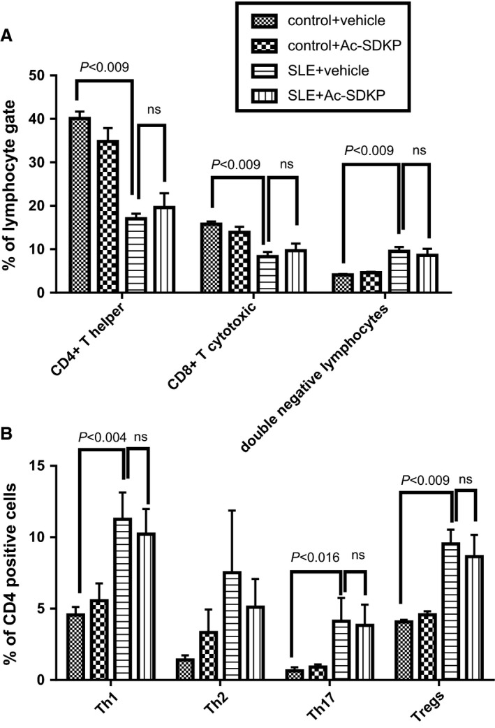
Figure 6.

Flow cytometry analysis of peripheral blood cells sampled at 32–33 weeks of age in control and SLE with hypertension mice treated either with vehicle or Ac‐SDKP (2400 μg/kg per day). (A) Percentage of CD4‐positive, CD8‐positive, and double negative lymphocytes. (B) Percentage of CD4‐positive Th1, Th2, T17 T cells, and Tregs. SLE, systemic lupus erythematosus; Ac‐SDKP, N‐acetyl‐seryl‐aspartyl‐lysyl‐proline.
