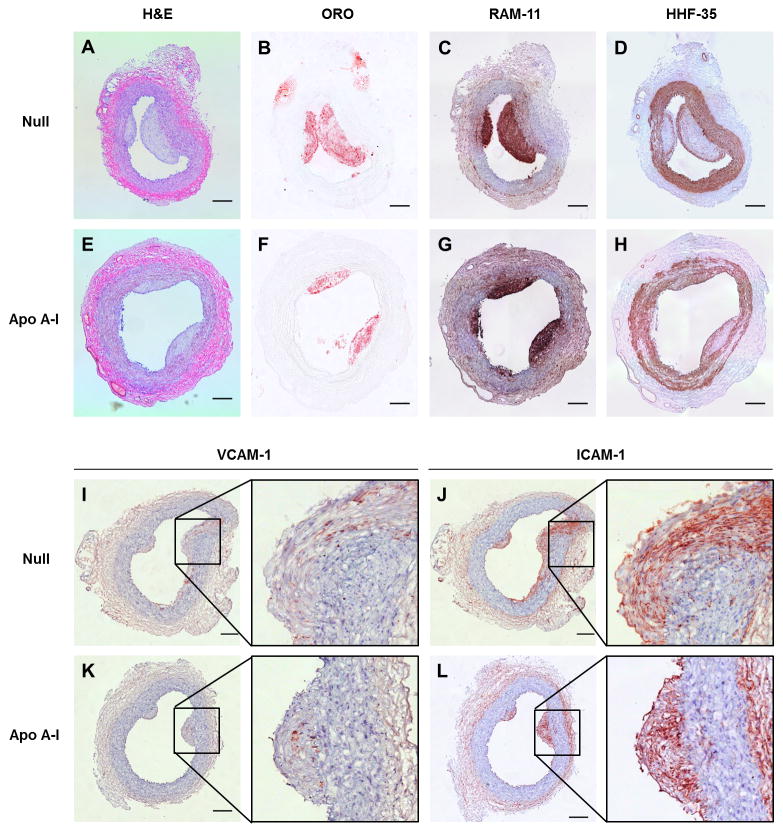
Figure 3.
Carotid atherosclerotic lesions 35 weeks after beginning high-fat diet and 7 weeks after infusion of HDAdNull or HDAdApoAI. Arteries were removed, embedded in OCT, and sectioned at multiple steps. Sections of representative lesions treated either with HDAdNull (Null) or HDAdApoAI (Apo A–I) are shown, stained with: hematoxylin and eosin (H & E) (A, E); Oil Red O (ORO) (B, F); RAM-11 antibody to detect macrophages (C, G); HHF-35 antibody to detect smooth muscle actin (D, H); anti-VCAM-1 (I, K); or anti-ICAM-1 (J, L). Sections A, B, C, and D are from the same artery, as are: sections E, F, G, and H; sections I and J; and sections K and L. All panels except B and F: hematoxylin counterstain. Scale bars = 200 μm.

