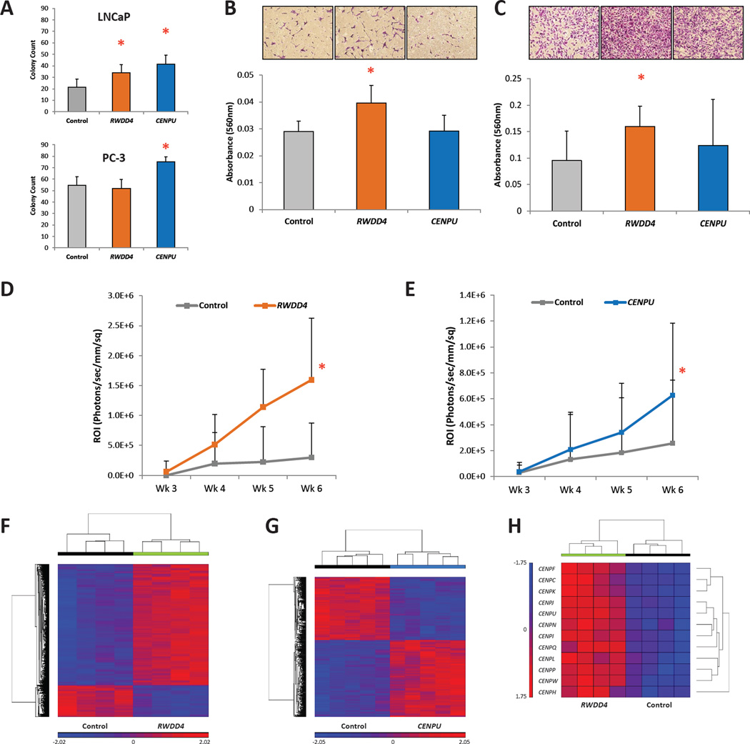
Figure 5.
In vitro and in vivo analysis of the effects of RWDD4 and CENPU dysregulation on LNCaP and PC-3 cells. (A) Soft agar anchorage-independent growth assays for LNCaP (upper panel) and PC-3 (lower panel) cells over-expressing either RWDD4 or CENPU (mean ± SD). (B) Trans-well invasion assay for LNCaP cells over-expressing either candidate gene (mean ± SD). (C) Trans-well invasion assay for PC-3 cells over-expressing either candidate gene (mean ± SD). (D) Intracardiac tumor dissemination assay for NU/J mice injected with either PC-3 cells over-expressing RWDD4 (n = 9) or a control cell line (n = 8) (mean ± SD). (E) Intracardiac tumor dissemination assay for NU/J mice injected with either PC-3 cells over-expressing CENPU (n = 20) or a control cell line (n = 16) (mean ± SD). (F) Microarray analysis of PC-3 cells over-expressing RWDD4 revealed dysregulation of 5,732 transcripts (fold change ± 1.5; FDR < 0.050). (G) Microarray analysis of PC-3 cells over-expressing CENPU revealed dysregulation of 682 transcripts (fold change ± 1.5; FDR < 0.050). (H) Multiple transcripts encoding centromere components are dysregulated in PC-3 cells over-expressing RWDD4. * P < 0.050. Also see Fig. S7.

