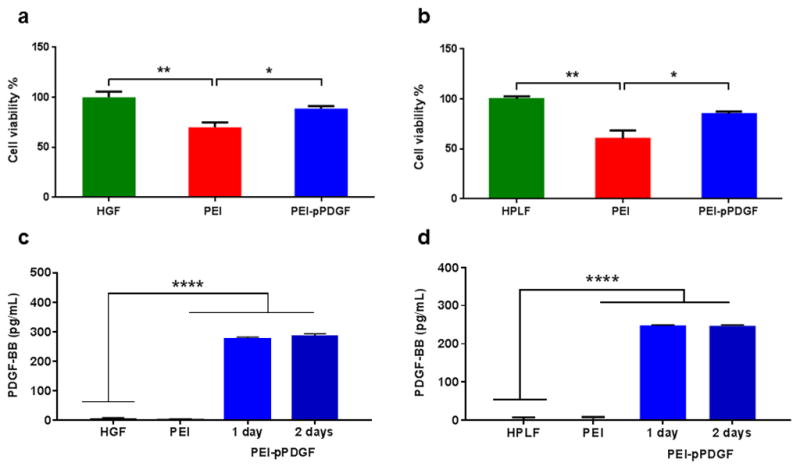
Figure 2.

Effects of PEI-pPDGF polyplexes fabricated at N/P ratio of 10 (1 μg pPDGF) on hGF (a) and hPLF (b) cells after 48 h. Significant differences between the treatments and the untreated cells were assessed by one-way analysis of variance followed by Tukey's post-test (*p<0.05; **p<0.01). Values are expressed as mean ± SD (n = 4). ELISA assay demonstrating the expression of PDGF-BB protein from hGF (c) and hPLF (d) cells at 24 and 48 hours, post transfection with PEI-pPDGF polyplexes (Prepared at an N/P ratio of 10 (1 μg pPDGF). Significant differences between PEI-pPDGF, cells treated with PEI and untreated cells were assessed by one-way analysis of variance followed by Tukey's post-test (****p < 0.0001). Values are expressed as mean ± SD (n = 4).
