Figure 1.

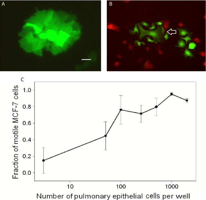
MCF‐7 cells transfected with GFP grown in standard culture conditions (A), and with SAEC labeled red with SNARF ®‐1 carboxylic acid, acetate succinimidyl ester (B). MCF‐7 cells separate from the clusters and display pseudopodia and lamellipodia (arrow). Original magnification 400×, scale bar = 50 μm. MCF‐7 scattering at different densities of SAEC (C), revealing dose‐dependent scattering with increasing numbers of SAEC cells. Error bars show standard errors of the mean. SAEC, small airway epithelial cells; GFP, green fluorescent protein.
