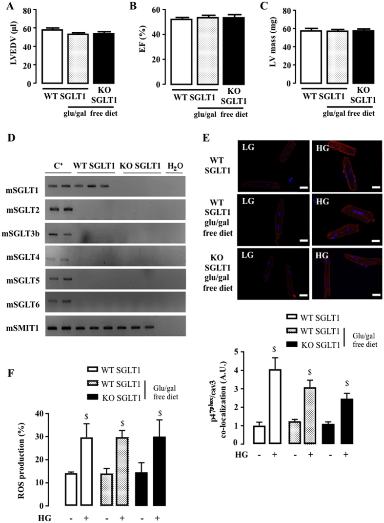
Figure 5. Impact of SGLT1 deletion on HG-induced NOX2 activation and ROS production.
(A) LVEDV, (B) EF and (C) LV mass were measured by echocardiography of SGLT1 WT under usual diet (n = 10), of SGLT1 WT submitted to glucose and galactose free diet (glu/gal free diet, n = 10) and of SGLT1 KO (n = 10) mice. Echocardiographic data in M-mode and 2D parasternal long axis are presented in Supplementary Table 4. (D) Detection of SGLT1, SGLT2, SGLT3b, SGLT4, SGLT5, SGLT6 and SMIT1 by RT-PCR and ethidium bromide-stained agarose gels on mRNA extracted from the hearts of SGLT1 KO mice (n = 3) compared to WT mice (n = 3). Positive controls were intestine for SGLT1, kidneys for SGLT2, SGLT3, SGLT4 and SGLT5, brain for SGLT6 and SMIT1. (E) Quantification of HG-induced p47phox translocation close to cav3 in SGLT1 WT mice (with and without glu/gal free diet) compared to SGLT1 KO mice. Adult mouse cardiomyocytes were isolated from SGLT1 WT (n = 7), SGLT1 WT submitted to glu/gal free diet (n = 7) and SGLT1 KO (n = 7) hearts. PLA was performed 90 min after stimulation with HG and compared to LG. White lines correspond to 20 μm. (F) ROS production induced by 3 h of incubation with HG in cardiomyocytes isolated from SGLT1 WT (n = 6), SGLT1 WT submitted to glu/gal free diet (n = 6) and SGLT1 KO (n = 6) mice. Data are means ± SEM. Statistical analysis was by two-way ANOVA. $Indicates values statistically different from LG, p ≤ 0.05.

