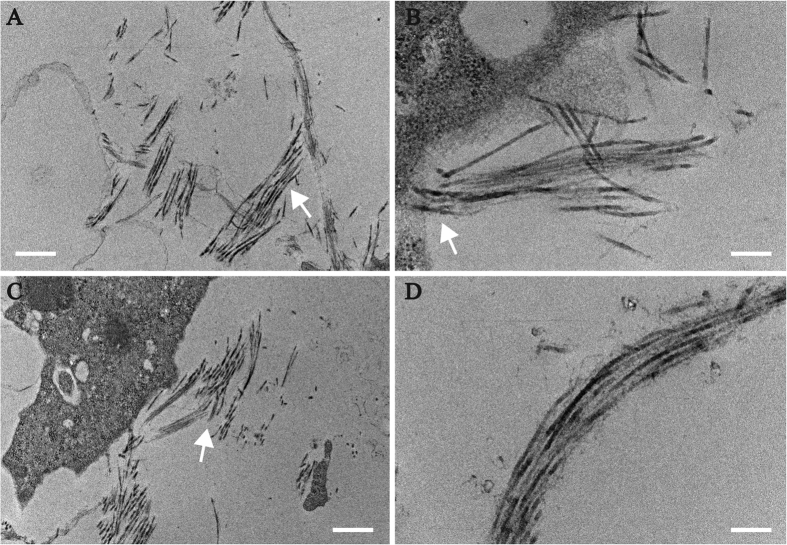
Figure 5. Collagen fibril accumulation in organoids.
TEM showing arrangement of presumptive collagen fibrils viewed surrounding the cells. Collagen fibrils are highlighted with arrows in (A–C) a presumptive packed lamella with stacked collagen fibril is shown in (D). Scale bars A-0.5 μm, B-200 nm, C-0.5 μm, D-200 nm.

