
Figure 6.
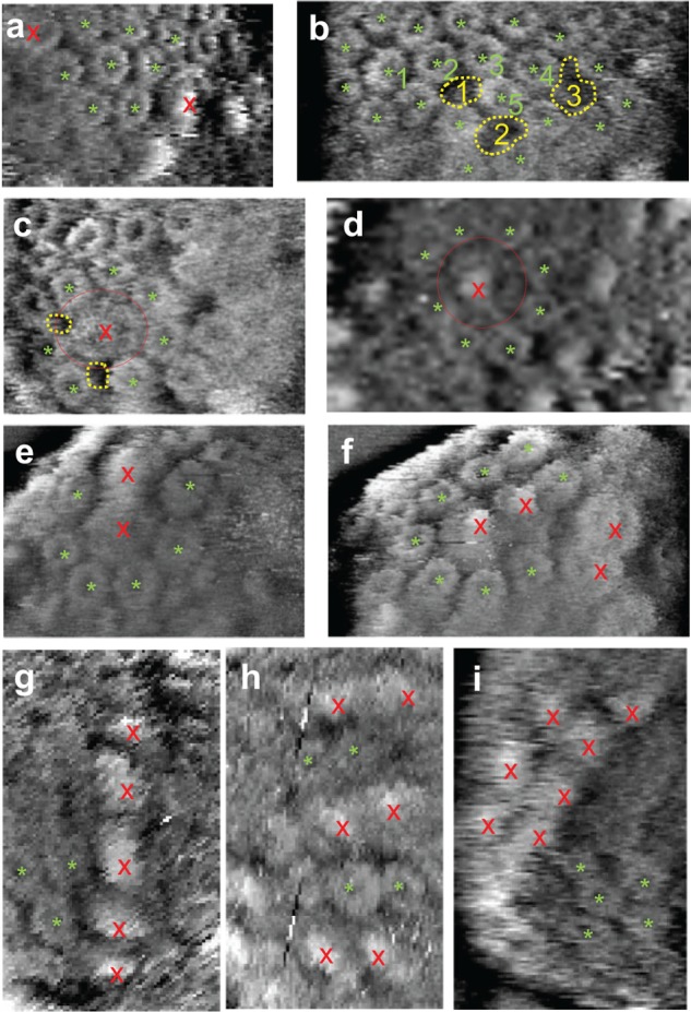
High-resolution AFM images showing example arrangements of LH2s and core complexes. (a) Hexagonal packing of LH2 rings (green *). Also in (a), there are three rows of LH2 rings between two core complexes (red ×). (b) LH2-rich region where there are some “empty lipid” regions (yellow dots). “Empty lipid” regions 1 and 2 are surrounded by five LH2 rings, while “empty lipid” region 3 is surrounded by six LH2 rings. LH2 ring 1 is surrounded by seven LH2 rings, LH2 rings 2, 3, and 4 are surrounded by five rings and an “empty lipid” region, while LH2 ring 5 is surrounded by four rings and two “empty lipid” regions. (c and d) Two monomer core complexes (red dotted circle) surrounded by LH2 rings. In (c) the core is surrounded by seven LH2 rings and “empty lipid” regions, while in (d) the core is surrounded by eight LH2 rings. (e and f) Two core complex dimers. In (e) the dimer is surrounded by LH2 rings and “empty lipid” regions, and by symmetry it can be said that 10 LH2 rings and “empty lipid” regions surround a core dimer. Also, (e) shows that each core of an S-dimer can be in contact with up to six LH2 rings. In (f) the dimer is surrounded by LH2 rings, “empty lipid” regions, and another core dimer. (g) Linear arrangement of five core complexes, and each core is in contact with at least four LH2 rings. (h) Alternate rows of core dimers and LH2 rings. (i) Seven core complexes in two rows next to each other with each core complex in contact with at least two LH2 rings. Some core complexes have a lower protrusion in the center, suggesting decapitation of parts of the RC complex, possibly due to the imaging force.