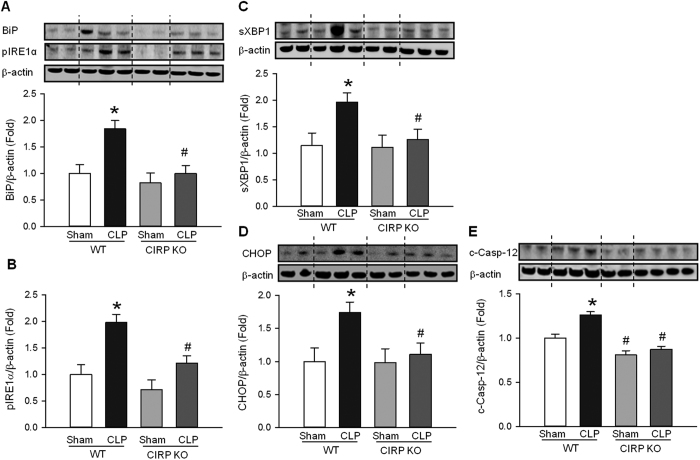
Figure 3. CIRP promotes ER stress during ALI.
Lung protein levels of (A) BiP, (B) phosphorylated IRE1α (pIRE1α), (C) spliced XBP1 (sXBP1), (D) CHOP and (E) cleaved caspase-12 (c-Casp-12) were determined by Western blotting (WB) in homogenates of lungs collected 20 h after CLP or sham operation. The levels of all five ER stress proteins were elevated in septic WT mice (*P < 0.05 vs. sham WT mice) and were significantly reduced in septic CIRP KO mice (#P < 0.05 vs. septic WT mice). The dotted lines on the WB reflect the groups shown in the histogram below. Histograms show mean densitometric analysis of bands normalized for β-actin and relative to expression values in sham WT mice. Data are expressed as last square mean ± SEM. Sample sizes: sham WT = 4, CLP WT = 6 sham CIRP KO = 4, CLP CIRP KO = 6.

