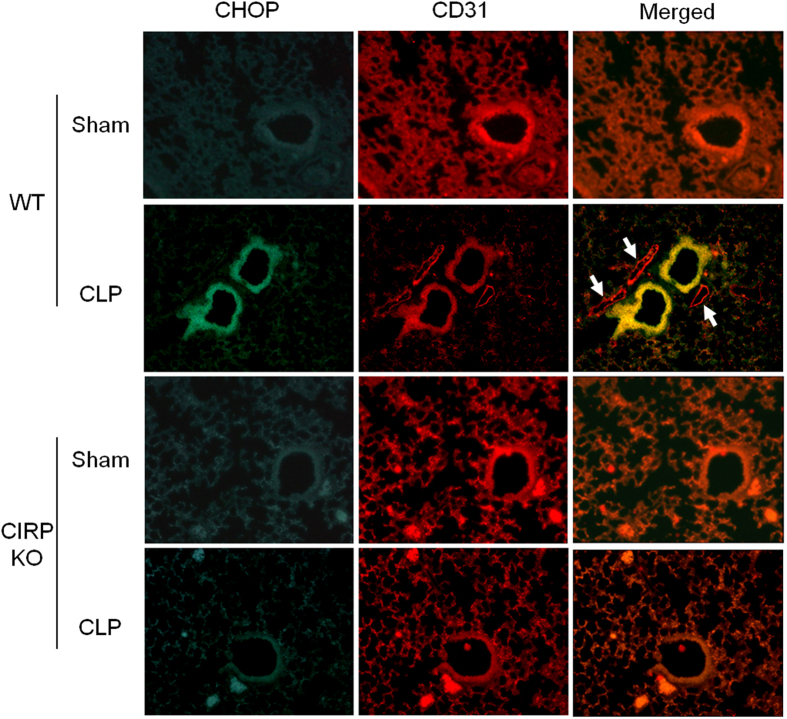
Figure 4. CIRP induces CHOP expression in pulmonary arteriolar and microvascular endothelial cells.
The expression of CHOP (green) and CD31 (red) was determined by immunofluorescence in lungs collected 20 h after CLP or sham operation. High expression of CHOP was detected in pulmonary arteriolar endothelial cells of septic WT mice, but not in the other groups. CHOP was highly expressed in arteriolar endothelial cells (merged, yellow), and to a lesser extent in microvascular endothelial cells (merged, orange). CHOP was not detected in venular endothelial cells of septic WT mice (merged, white arrows). Representative sections; original magnification, 200X.

