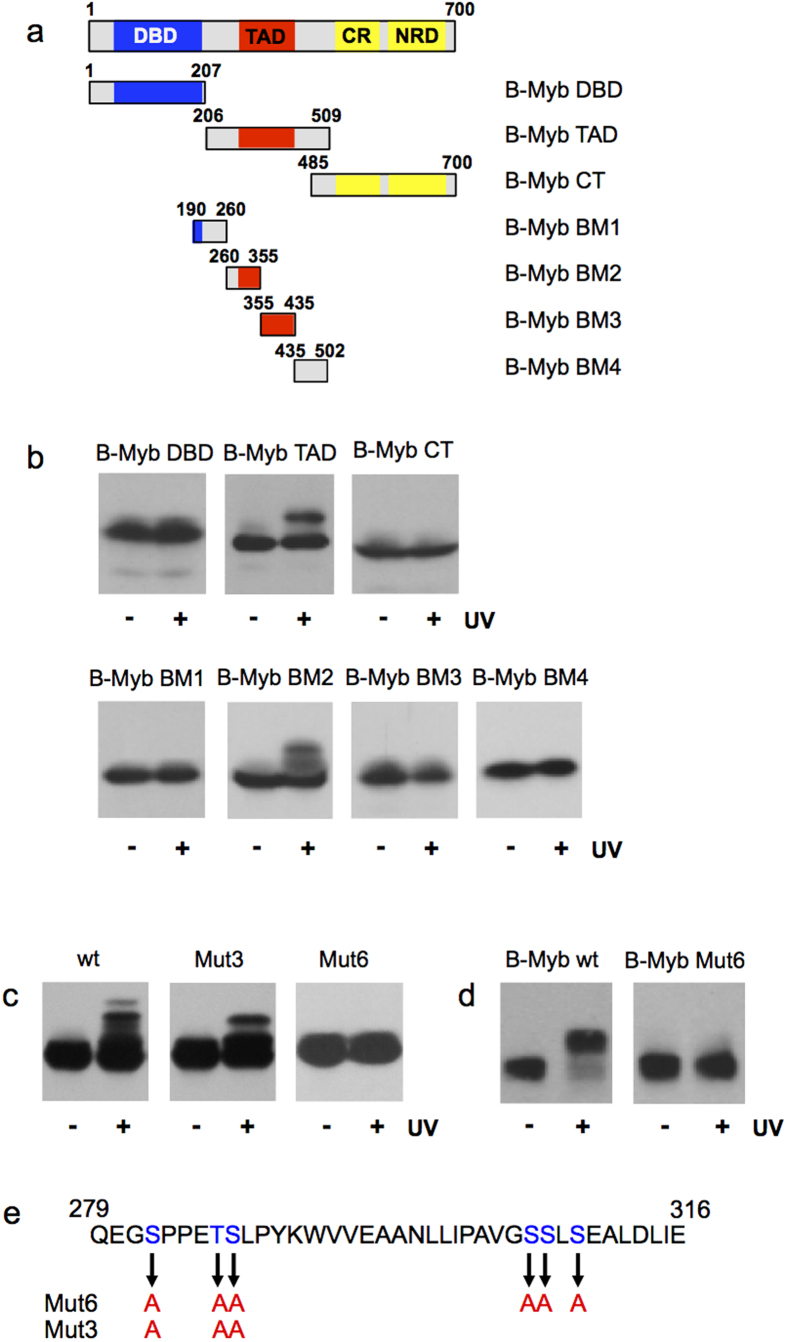
Figure 5. Mapping of the sites of UV-induced phosphorylation of B-Myb.
(a) GFP/B-Myb constructs are shown schematically at the top. (b) The indicated B-Myb constructs were expressed in QT6 fibroblasts prelabeled with BrdU. The cells were UV irradiated (30 J/m2) and harvested after 60 min. Unirradiated cells served as controls. Total cell extracts were analyzed by western blotting with antibodies against GFP (bottom panels). (c) Wild-type B-Myb BM2 and mutants Mut3 and Mut6 were analyzed as in panel B. (d) Full-length B-Myb-wt and B-Myb-Mut6 were analyzed as in panel B. (e) Overview of amino acid residues changed to Ala in the B-Myb mutants.

