
Figure 1. NMD proteins are increased after SE in mice and Upf1 is increased in human TLE samples.
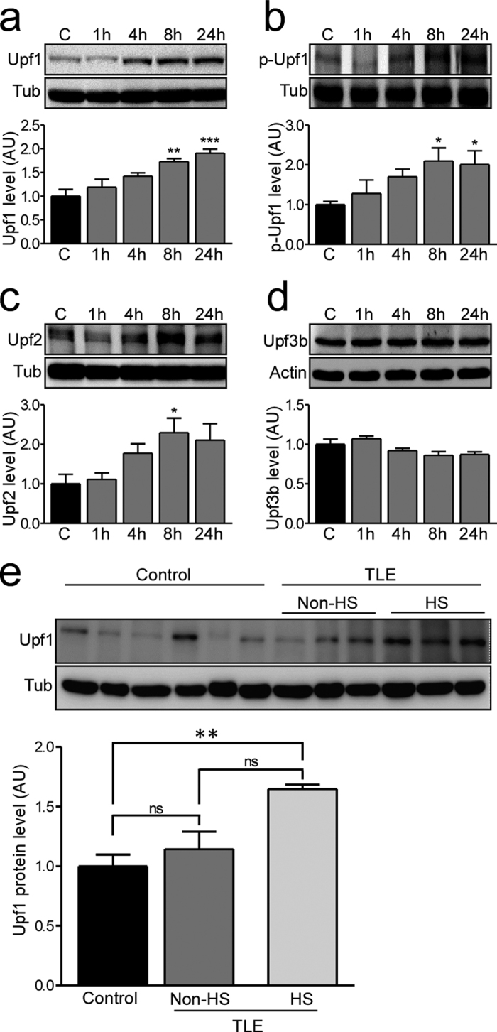
(a–d) Protein levels of Upf1, phosho-Upf1 (p-Upf1), Upf2 and Upf3b in the ipsilateral hippocampus in control (C) mice and at 1, 4, 8 and 24 hours (h) after SE were analysed by western blot and semi-quantified. (a) Upf1 levels significantly increased 8 and 24 h after SE (n = 4/group; ANOVA, Dunnett’s posthoc test ***p < 0.001, **p < 0.01). (b) p-Upf1 levels significantly increased 8 and 24 h after SE (n = 5/group; ANOVA, Dunnett’s posthoc test *p < 0.05 compared to control. (c) Upf2 levels were increased 8 h after SE (n = 4/group; ANOVA, Dunnett’s posthoc test *p < 0.05 compared to control). (d) Upf3b levels did not change after SE (n = 4/group; ANOVA, Dunnett’s posthoc test p = 0.88). (e) Upf1 protein levels were significantly higher in TLE patients with hippocampal sclerosis (HS) compared to post-mortem controls and TLE patients without HS (Controls n = 6; TLE without HS n = 3, TLE with HS n = 3; ANOVA with Bonferroni post-hoc test comparing all columns; p = 0.0072). Representative blots have been cropped to reduce unnecessary area.