
Figure 2. Hippocampal re-localization of Upf1 after SE in mice.
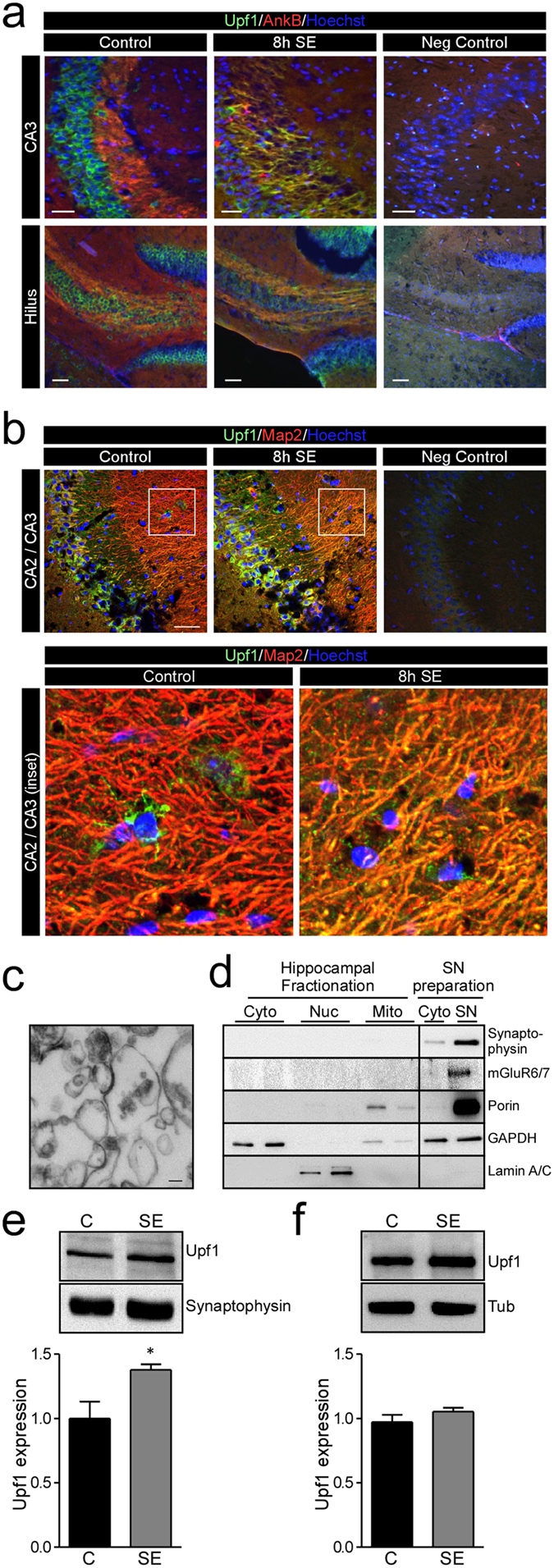
(a, b) Representative photomicrographs of mouse hippocampal sections stained with antibodies against Upf1 (green), dendrite marker protein Map2 (red) or axon marker protein AnkyrinB (AnkB, red) and Hoechst (blue). Negative control sections were prepared with no primary antibodies. Images show increased Upf1 colocalisation with AnkB in the CA3 and hilus subfields (a) and increased colocalisation with Map2 in the CA2/CA3 regions (b) 8 h after SE. (c) Mouse synaptoneurosomes (SN) were prepared and synaptic enrichment was confirmed using electron microscopy by the presence of morphological features of synaptoneurosome including a dark post-synaptic density between a synapsome and synaptoneurosome. (d) Western blot analysis confirming enrichment of synaptic proteins synaptophysin and mGluR6/7 and mitochondrial protein porin. (e) Western blot analysis of Upf1 levels show Upf1 was increased in synaptoneurosomes 8 h after SE in mice (Control n = 4, SE n = 5; student’s t-test p = 0.019). (f) SE did not significantly alter Upf1 levels in the corresponding cytoplasmic fraction (Control n = 4, SE n = 5; student’s t-test p = 0.2130). Representative blots have been cropped to reduce unnecessary area.