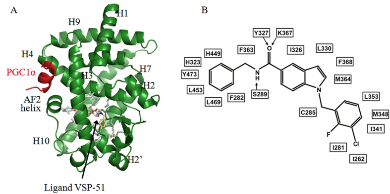
Figure 6. Crystal structure of PPARγ LBD in complex with VSP-51.
(A) The overall PPARγ LBD structure (green) in complex with compound VSP-51 (pink) and a PGC1α peptide (red). Labeled are major secondary structure features, and the compound. The residues surrounding the compound are presented as sticks with carbons in grey, nitrogens in blue, and oxygens in red. (B) A schematic presentation of the interaction network between compound VSP-51 and the pocket residues of PPARγ. Arrows indicate H-bonds.

