
Fig. 3.
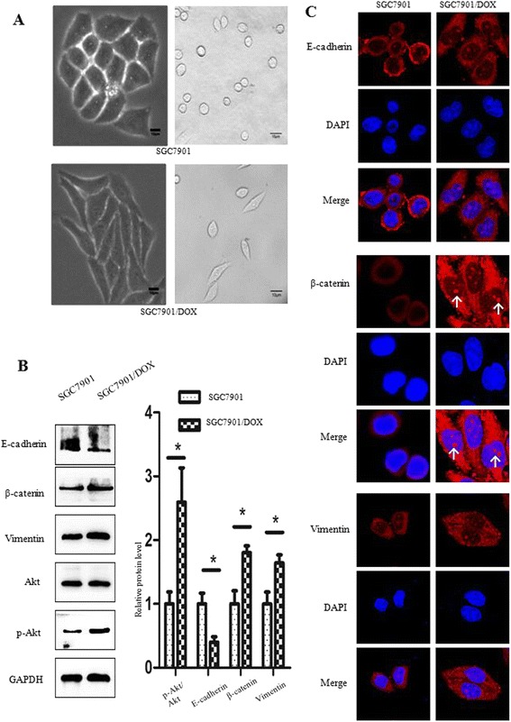
SGC7901/DOX cells underwent EMT mediated by upregulation of Akt signaling pathway. a Images were captured by Olympus IX71 inverted microscope system. Morphological observation showed morphological variance between SGC7901/DOX and SGC7901 cells. The Scale bar represents 10 μm. b SGC7901 and SGC7901/DOX cells were left DOX-untreated for 48 h for Western blot assay, measeuring EMT-related proteins and Akt expression. Bar diagram shows the relative expressions of proteins normalized to GAPDH. Data are represented as mean ± SD of three independent experiments (n = 3, *** p < 0.001; ** p < 0.01; * p < 0.05: NS means not significant, p > 0.05). c Immunofluorescence assay was used to detect EMT-associated proteins in SGC7901/DOX cells. E-cadherin, β-catenin, and vimentin were stained red and nuclei stained with DAPI were blue. The white arrow indicates β-catenin nuclear tranlocation. Images were captured at 1800× magnification