
Fig. 1.
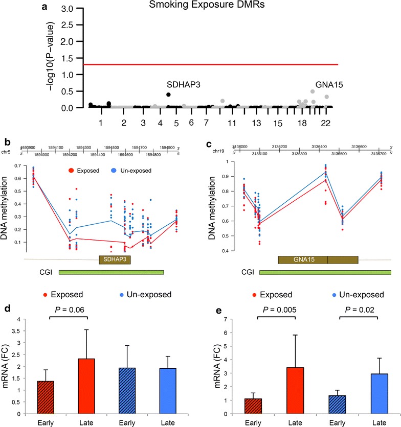
Differentially methylated regions associated with fetal smoking exposure. a Manhattan plot shows the most significant smoking exposure DMRs identified between smoking-exposed and unexposed fetal cortical samples, red line; adjusted p value = 0.05. DNA hypomethylation of fetal cortical samples exposed to maternal smoking was found within the promoter regions of the two most significant smoking exposure DMRs b SDHAP3 and c GNA15. CGI CpG Island. Gene expression of d SDHAP3 and e GNA15 shows temporal up-regulation in the fetal cortex, particularly in smoking exposed