
Fig. 3.
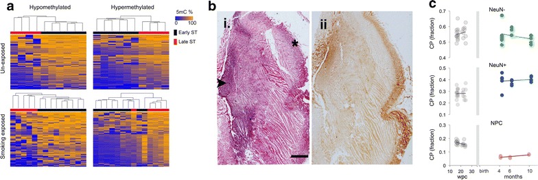
DNA methylation patterns of the developing fetal DLPFC. a Heatmaps of unsupervised hierarchical clustered DMPs found differentially methylated between early and late ST for each exposed and unexposed group. b Coronal sections from formalin-fixed, paraffin-embedded, previously frozen fetal cortex (20 wpc). Frontal region of cerebral hemisphere showing (i) hematoxylin and eosin (H&E) staining and (ii) NeuN staining, which distinctly labels the cortical plate (asterisk on H&E) and germinal matrix (arrowhead) (Scale = 2 mm). c CP estimates of NeuN−, NeuN+ and NPC within exposed and unexposed (local) fetal DLPFC samples (gray) were compared to CP estimates of publicly available postnatal frontal cortex generated by BrainSpan consortium (colored) (“Methods” section)