Fig. 1.

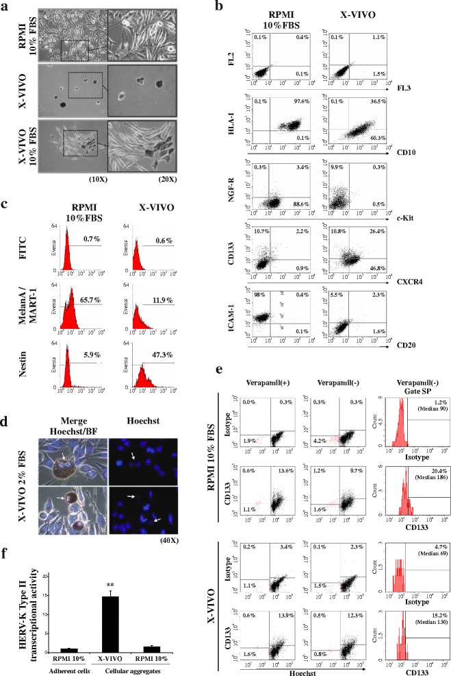
TVM-A12 cells exhibit adaptive plasticity, phenotype switching and concomitant up-regulation of HERV-K expression upon microenvironmental modifications. a Phase contrast microscopy of TVM-A12 grown in RPMI-1640 complete medium, in stem cell medium (X-VIVO) or upon the addition of increasing percentage of serum (up to 10% of FBS), where cells reverse their original phenotype within 72 h; original magnification: left panels, 10x; right panels, 20x. b Flow cytometry analysis after extracellular staining of TVM-A12 melanoma cells undergoing from adherent growth, in RPMI-1640 complete medium, to grape-like cellular aggregates in X-VIVO medium. c Flow cytometry analysis after intracellular staining of TVM-A12 melanoma cells undergoing from adherent growth, in RPMI-1640 complete medium, to grape-like cellular aggregates in X-VIVO medium. d Fluorescence microscopic analysis of Hoechst 33342 staining on living TVM-A12 cells growing in low serum X-VIVO medium (2% FBS); arrows point to putative cancer stem cells exhibiting low or negative Hoechst positivity in the nuclei. Original magnification: 40x. e Shows identification and characterization of the SP cells from TVM-A12 melanoma cells cultured for 72 h in RPMI-1640 complete medium or X-VIVO using Hoechst 33342 nucleic acid stain by FACS analysis. The FACS dot-plots displays the SP cells co-stained with CD133 marker preincubated with verapamil (left panel) and in the absence of verapamil (middle panel). Likewise, the FACS histograms plots (right panel) displays the percentage of CD133+ cells and the median of the relative fluorescence within the total SP cells in the absence of verapamil. f Relative mRNA expression of HERV-K env gene in TVM-A12 melanoma cells upon modification of culture conditions (** p < 0.001). Data represent the results of three independent experiments
