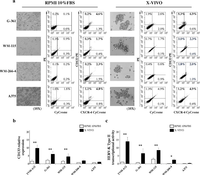
Fig. 4.

Different melanoma cell lines increase CD133 marker upon activation of HERV-K. a Morphological transition (left panel) and CD133 expression by flow cytometry (right panel) of human melanoma cell lines (G-361, WM-155, WM-266-4 and A375) upon exposure to X-VIVO medium: magnification 10x. Relative mRNA expression of CD133 (b) and HERV-K (c) in different melanoma cell lines upon modification of culture conditions. p-values (* p ≤ 0.050; ** p < 0.001). Data represent the results of three independent experiments
