FIGURE 3.
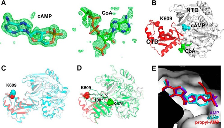
Crystal structure of SeAcs in complex with cAMP and CoA. A, simulated-annealing omit Fo − Fc electron density map (green) contoured at 3.0 σ for cAMP (left) and CoA (right) molecules. Blue, orange, and red represent nitrogen, phosphorus, and oxygen atoms, respectively. Cyan and green represent carbon atoms of cAMP and CoA, respectively. B, overall structure of SeAcs in complex with cAMP (cyan) and CoA (green). White and red ribbons represent the N-terminal and C-terminal domains of SeAcs, respectively; red sphere, Lys609; cyan sphere, cAMP; green sphere, CoA. C, superimposition of our SeAcs-cAMP-CoA crystal structure (red and gray) with the crystal structure of SeAcs complexed with AMP, CoA, and acetate (PDB code 2P2F; cyan). D, superimposition of our SeAcs-cAMP-CoA crystal structure (red and gray) with the crystal structure of yeast Acs in complex with AMP (PDB code 1RY2; green). E, close-up view of the cAMP (cyan) binding pocket with superimposed propyl-AMP (red) from the crystal structure of SeAcs complexed with propyl-AMP, CoA, and acetate (PDB code 1PG4), and with superimposed AMP (purple) from the crystal structure of SeAcs in complex with AMP (PDB code 2P2F).

