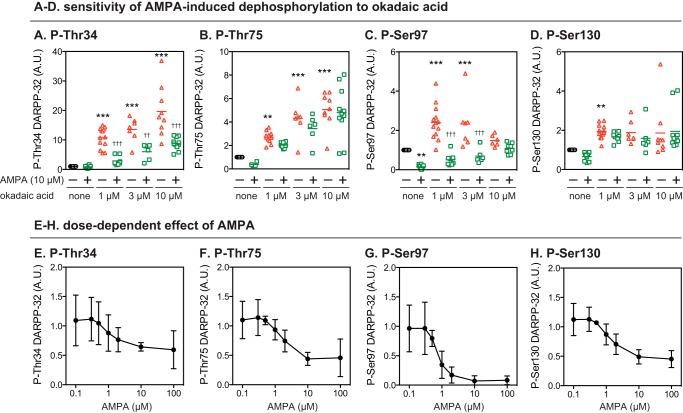
FIGURE 3.
Effects of okadaic acid on AMPA-induced dephosphorylation of DARPP-32 at four phosphorylation sites. A–D, striatal slices were incubated with okadaic acid at 1, 3, or 10 μm for a total of 70 min, with the addition of AMPA (10 μm) for the final 10 min. Phosphorylation of DARPP-32 at the four indicated sites was determined by immunoblotting as in Fig. 1. Data for 6–13 experiments are presented in scatter plots with the mean values indicated by a line. **, p < 0.01; ***, p < 0.001 compared with untreated slices; ††, p < 0.01; †††, p < 0.001 compared with okadaic acid alone at the same concentrations; one-way ANOVA followed by Newman-Keuls test. E–H, striatal slices were treated with AMPA at various concentrations (0.1–100 μm) for 10 min. Phosphorylation of DARPP-32 at the four indicated sites was determined by immunoblotting. Data represent the means ± S.D. for 3–12 experiments. A.U., arbitrary units.

