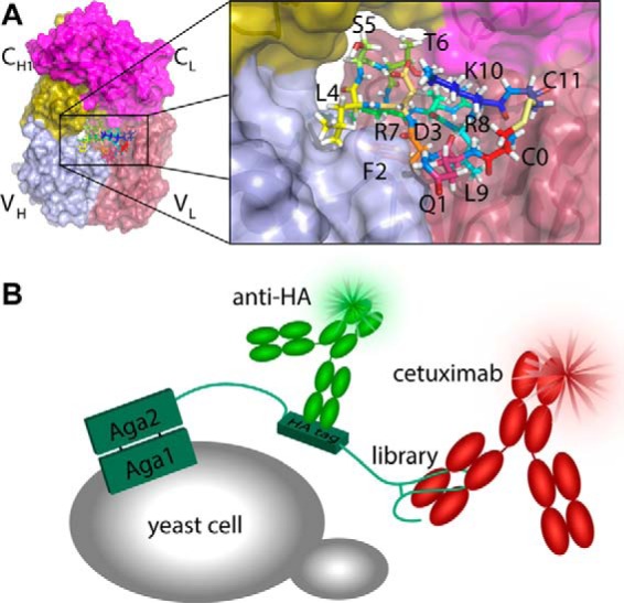
FIGURE 1.

Improving cyclic peptide binding to cetuximab using yeast display. A, structure of the Fab fragment of cetuximab indicating the position of the meditope in the central cavity. B, schematic representation of the yeast display system. Simultaneous labeling of the HA tag and cetuximab with different fluorophores allows selection for binding to be normalized for variations in expression.
