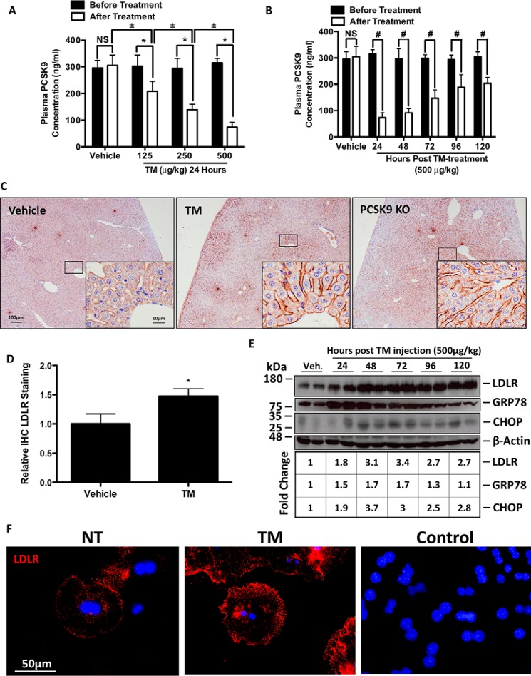
FIGURE 5.
TM blocks PCSK9 secretion and induces hepatic cell surface LDLR expression in vivo. 12-Week-old C57BL/6 female mice were randomly divided into eight groups (n = 3) and subcutaneously injected with a single dose of PBS vehicle control or TM. Animals treated with PBS or TM (125–500 μg/kg) were sacrificed 24 h following injection. The remaining animals, belonging to groups 5–8 were treated with a single injection of TM (500 μg/kg) and sacrificed every 24 h for the following 120 h. Plasma was taken from each mouse prior to injection and after sacrifice. A, *, p < 0.05 versus before treatment. ±, p < 0.05 versus after treatment. A and B, plasma PCSK9 levels were quantified via ELISAs. B, #, p < 0.05 versus before treatment. C and D, livers from the TM-treated and untreated PCSK9 KO mice were collected, fixed in formalin, sectioned, stained for LDLR, and quantified. E, flash-frozen liver tissues from these animals were used to examine the expression of LDLR and of UPR markers GRP78 and CHOP using immunoblots. *, p < 0.05 versus vehicle. F, to elucidate whether IHC LDLR stating occurred specifically on cell surface LDLR, mouse primary hepatocytes were plated and stained for LDLR in the absence of permeabilizing agents. Control, HuH7 cells were also incubated with Alexa 594 secondary antibody, in the absence of primary antibody staining. Differences between treatments were assessed with unpaired Student's t tests, and all values are represented as mean ± S.D. Veh, vehicle; NS, not significant.

