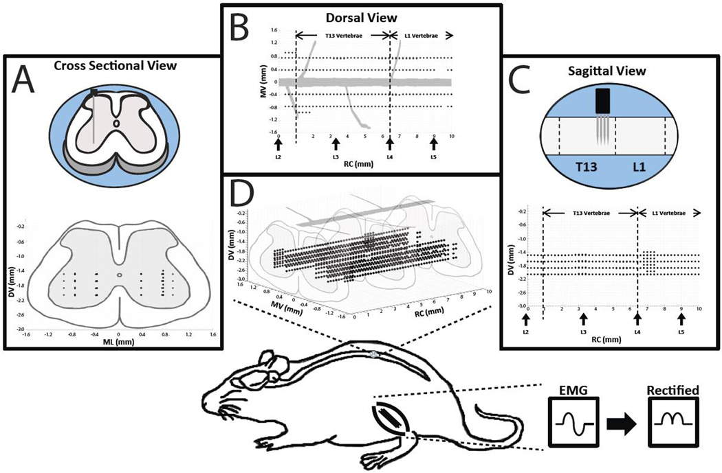
Figure 2. Overview of Experimental Design.
Intraspinal microstimulation was conducted in the T13-L1 vertebral segments (L2-S1 spinal segments) in nine rats. Stimulation sites (black dots) were located in rostrocaudal (RC; 0 – 10 mm from the L2 nerve root), mediolateral (ML; 0.4 and 0.8 mm from midline), and dorsoventral (DV; 1.6 – 2.2 mm below the surface of the spinal cord) dimensions. A) A cross sectional view of the rat spinal cord showing the dorsoventral (DV) and mediolateral (ML) locations of the stimulation sites while collapsing the rostrocaudal (RC) locations. B) A dorsal view of the rat spinal cord showing the ML and RC locations of the stimulation sites while collapsing the DV locations. Vertical arrows on the x-axis indicate the approximate locations of the dorsal roots. C) A sagittal view of the rat spinal cord showing the DV and RC locations of the stimulation sites while collapsing the ML locations. D) The three dimensional topographic map derived from the cross sectional, dorsal, and sagittal views. In one rat, stimulation sites were explored in a third ML track (1.0 mm from midline). Bottom: Location of the lumbar cord examined in the rat. In three rats, EMG signals from selected hindlimb muscles were recorded during ISMS, filtered, full-wave rectified, and averaged during each stimulation trial.

