Figure 3.

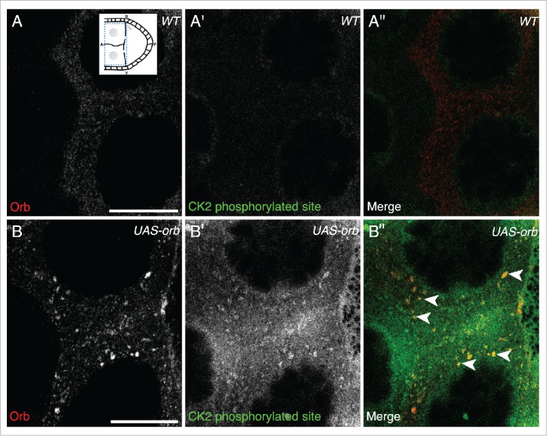
In UAS-Orb chamber, ectopically expressed Orb is phosphorylated by CK2. Double immunofluorescent staining of wild type (A-A″) and tubGal4-VP16; UAS-Orb (B-B″) egg chambers for Orb protein (red) and phosphorylated CK2 consensus sites (pS/pT-D-X-E, green). Inset: schematic illustrating the relative position of the oocyte and nurse cells for a stage 8 egg chamber, dashed blue box highlights area focused on in both (A-A″) and (B-B″). (A-A″) In the nurse cells of wild-type egg chambers, Orb protein is at a low level and CK2 activity is below the level of detection (n = 12/12). (B) tubGal4-VP16; UAS-Orb egg chambers, have ectopically expressed Orb in foci of varying sizes throughout the nurse cells (n=12/12). (B′) Phosphorylated CK2 consensus sites are visible in tubGal4-VP16; UAS-Orb egg chambers (n = 12/12). (B″) Merge of 2 stains showing that ectopically expressed Orb protein in the nurse cells is phosphorylated by CK2 (white arrowheads). Single slices, scale bar 20 μm.
