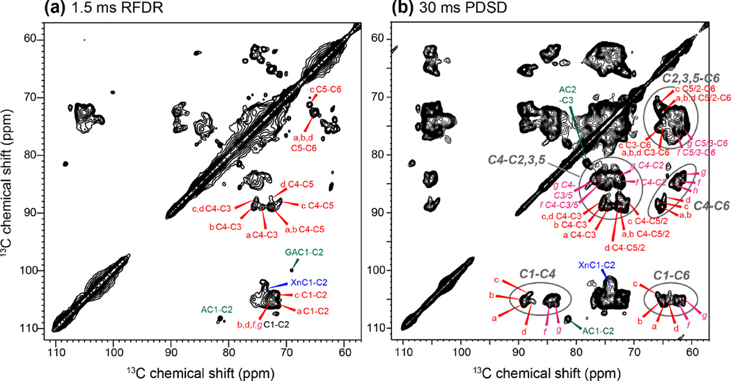
Figure 2.
2D 13C-13C correlation spectra of Brachypodium primary cell walls for resonance assignment of the polymorphic cellulose. (a) 1.5 ms RFDR spectrum showing one-bond cross peaks. (b) 30 ms PDSD spectrum showing intra-residue multi-bond cross peaks. The interior and surface cellulose signals are annotated in red and magenta, respectively. Ovals guide the eye for characteristic cross peak regions such as C1-C4, C4-C6 and C1-C6.

