Figure 6.
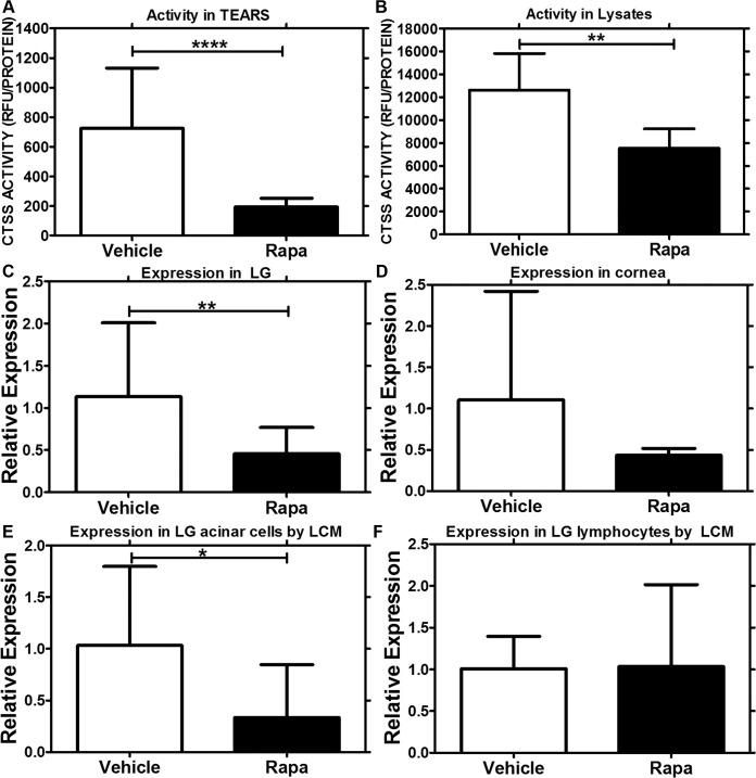
Decrease in CTSS activity and gene expression in tears, LG lysates, cornea, and LG acinar cells from mice treated with Rapa eye drops. (A) Tear CTSS activity measurements indicated a 6-fold decrease (P < 0.0001) in mice treated with Rapa eye drops (n = 9), as compared with those treated with vehicle alone (n = 10). (B) Cathepsin S activity measurements in LG lysates also demonstrated a significant decrease (P = 0.003) in mice treated with Rapa (n = 6), as compared with those treated with vehicle (n = 5). (C) Quantitative (real-time) PCR studies demonstrated that the CTSS mRNA level was significantly lower in whole LG (P = 0.008) in mice treated with Rapa (n = 5), as compared with those treated with vehicle alone (n = 5). (D) Although we saw a trend in CTSS reduction in cornea of mice treated with Rapa (n = 3), it was not statistically significant (P = 0.15). To determine if CTSS expression was modified in LG acinar cells or invading lymphocytes, we used laser capture microdissection to isolate individual acini and lymphocytes from hematoxylin-stained LG sections of Rapa- (n = 3) and vehicle- (n = 3) treated mice and carried out qPCR on these cell types. As can be seen in (E), there was a significant reduction in CTSS mRNA expression in the LG acinar cells with Rapa treatment when compared with the vehicle treatment group (P = 0.04); (F) CTSS mRNA expression was unchanged in the lymphocytes. Data are presented as mean ± 95% CI.

