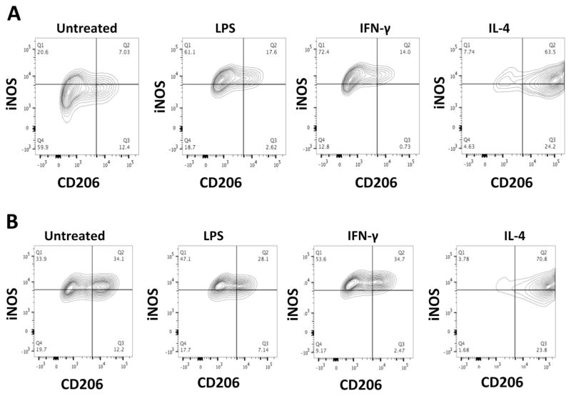
Fig. 1.
Characterization of mouse bone marrow-derived macrophages (a young; b aged) after polarization. Most of the aged M0s are both iNOS+ and CD206+ where as most of the young M0s are iNOS− and CD206−. There was no difference between young and aged M2 polarized cells and no difference between LPS and IFN-γ induced M1 polarization

