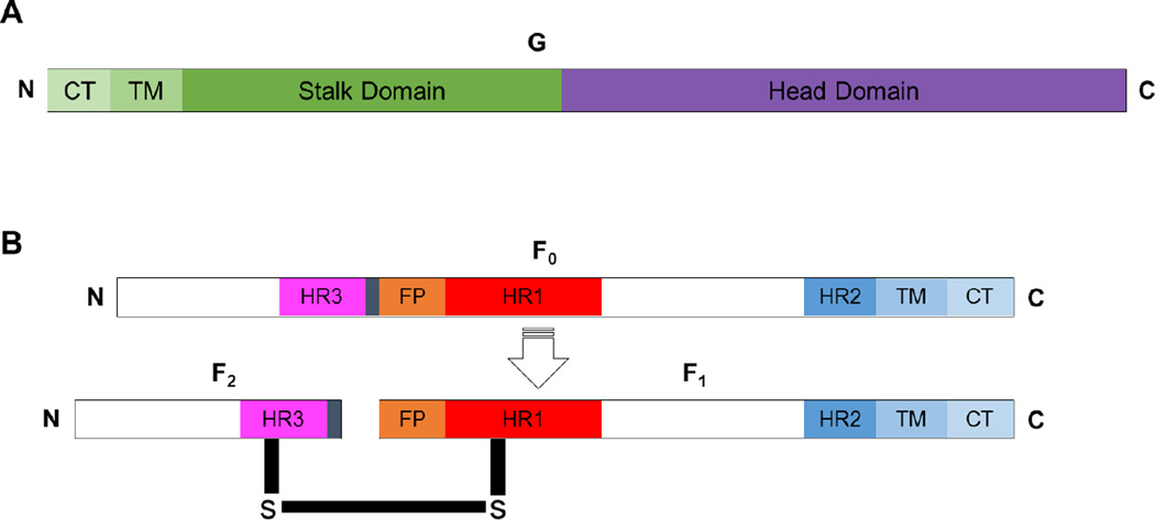
Fig. 2. Paramyxovirus attachment and fusion glycoproteins.
A) Diagram depicting the attachment glycoprotein. B) Diagram depicting the precursor of the paramyxovirus fusion glycoprotein (F0, top) and the cleaved, biologically active and disulfide linked paramyxovirus protein (F1-F2, bottom). The fusion peptide (FP), heptad repeats HR1, HR2, and HR3, transmembrane (TM); and cytoplasmic tail (CT) domains are shown, and the N- and C-termini are indicated.

