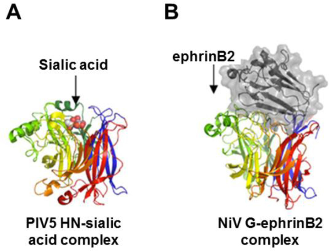
Fig. 3. Receptor binding with attachment glycoproteins occurs at the head domain.
The attachment protein binds the cellular receptor within a binding pocket(s) in the globular head. As illustrated, the glycoprotein monomer heads have separate β-blades distinguished by different colors for structures of the PIV5 HN (A) or NiV G (B). In both of these ribbon models sialic acid (A; orange cluster) or ephrinB2 (B; colored grey) binds the attachment protein towards the center of the β-propeller. This figure was adapted from (123). The PDB code for the NiV G-ephrin B2 complex is 2VSM (52).

