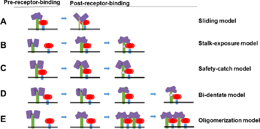
Fig. 5. Five models of F activation (A–E).
Attachment glycoprotein tetramers are colored in purple (heads) and green (stalks, transmembrane, and cytoplasmic tail domains). F is highlighted in red (heads) and blue (stalks, transmembrane, and cytoplasmic tail domains). In the bi-dentate model, the conformational changes of the head are shown as slightly distinct shapes. F-triggering regions in the stalk are shown as black lines (B–D).

