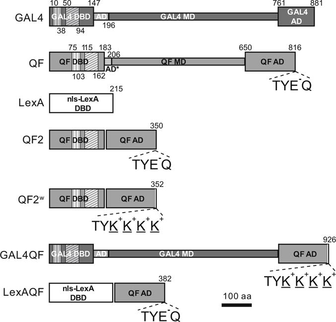
Fig. 3.

Transactivator schematics. Schematic representations of GAL4, original QF, LexA, QF2, QF2w, GAL4QF, and LexAQF. The transactivators consist of modular regions: DNA binding domain (DBD), middle domain (MD), and activation domain (AD). Vertical hatching indicates Zn 2/Cys 6 zinc finger motifs, diagonal hatchings mark dimerization domains. Numbers above and below schemes indicate amino acid position. Constructs are drawn to scale. C-terminal amino acids are indicated for transactivators with the QF AD to highlight differences between the ADs of QF/QF2/LexAQF and QF2w/GAL4QF
