
Fig. 5.
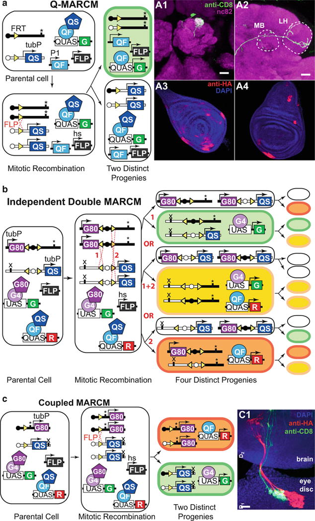
MARCM analysis. (a) Q-MARCM. The genotype of the parental cell is: hsFLP, FRT site (homozygous) recombined with tubulin-QS (heterozygous), P1-QF, QUAS-GFP. “*” marks the location of a recessive mutation that may be studied in the labeled cells. Upon FLPase-mediated mitotic recombination, one of the two postmitotic cells will lose the tubulin-QS transgene and start expressing GFP (top). The other postmitotic cell will remain unlabeled (bottom). A1 and A2: Q-MARCM labeling of a single olfactory projection neuron, visualized in the antennal lobe (A1), the mushroom body (MB, A2), and the lateral horn (LH, A2). Genotype: hsFLP, UAS-mCD8-GFP (X); GH146-QF#53, 82BFRT, tub-QS/82BFRT (III). A3 and A4: Q-MARCM labeled clones in the leg (A3) and wing (A4) imaginal disks of a third instar larva. Genotype: hsFLP1, QUAS-mtdT-3xHA (X); ET40-QF (II); 82BFRT, tubP-QS/82BFRT (III). Schematic and brain images reprinted with permission from ref. 15. (b) Independent double MARCM. The genotype of the parental cell is: hsFLP (also present in all progeny cells), P1-QF, P2-GAL4, UAS-GFP, QUAS-RFP, FRT site (homozygous) recombined with tubulin-GAL80 (heterozygous), a different FRT site (homozygous) recombined with tubulin-QS (heterozygous). “*” and “x” mark independent recessive mutations that may be studied in postmitotic cells. There are three possible outcomes of a heatshock-induced mitotic recombination at the FRT sites (1 or 2 or 1 + 2). The progenitors for each event are schematized with each generating a labeled and an unlabeled cell. Upon a second heatshock, mitotic recombination may happen again, altering the expression profiles of the progeny. See main text for details. Schematic modified with permission from ref. 15. (c) Coupled MARCM. The genotype of the parental cell is: hsFLP (also present in all progeny cells), P1-GAL4, P1-QF, UAS-GFP, QUAS-RFP, FRT site (homozygous) recombined with tubulin-GAL80 (heterozygous) or with tubulin-QS (heterozygous). “*” and “x” mark independent recessive mutations that may be studied in postmitotic cells. FLP-mediated recombination during mitosis at the FRT site followed by chromosome segregation result in all progeny being labeled either with GFP or with RFP. C1: Coupled MARCM clones in the eye-antennal imaginal disk send processes that innervate the brain of a third-instar larva. Genotype: hsFLP1, QUAS-mtdT-3xHA, UAS-mCD8-GFP (X); ET40-QF (II); 82BFRT tubP-QS/tubP-GAL4 82BFRT tubP-GAL80 (III). Schematic and image reprinted with permission from ref. 15