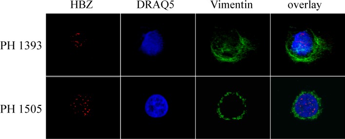
Fig 2. Subcellular localization of endogenous HBZ and Tax-1 in PBMC of ATL patients.
PBMC of two ATL patients (PH1393 and PH1505) were stained with the anti-HBZ 4D4-F3 mAb, followed by Alexa Fluor 546-conjugated goat anti-mouse IgG1antibody to detect the HBZ protein and analyzed by confocal microscopy. Specific counterstaining of nucleus or cytoplasmic compartments was performed by using DRAQ5 fluorescence probe or anti-vimentin antibody as describe in the legend to Fig 1. At least 300 cells were analyzed; a representative cell for HBZ staining is shown for each patient. PBMC from both patients were negative for expression of Tax-1 protein.

