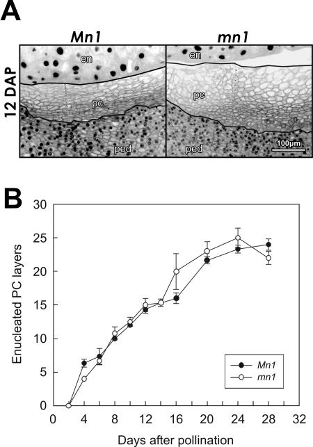
Figure 2.
Cellular morphology of maternal P-C cells lacking nuclei is determined by the filial endosperm. A, DAPI-stained longitudinal sections of Mn1 and mn1 maize caryopses at 12 DAP. Manually drawn lines bracket the cells without visible nuclei in the P-C layer and the crosses (×) inside the stack of cells demonstrate how the counting of cell layers was done. B, Temporal changes in the number of P-C layers without nuclei in Mn1 and mn1 caryopses. Mean ± sd of number of layers in median longitudinal sections of three different caryopses is shown. en, endosperm; pc, P-C layer; ped, pedicel.

